
Lumbar Pedicle Screw Fixation (CD Horizon® Sextant®)

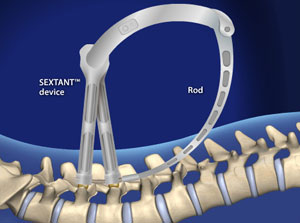
This minimally invasive procedure uses special guides and live x-ray imaging so the surgeon can place stabilizing screws and rods in the spine precisely, with less disruption to the muscles, tendons, and other soft tissue of the back.
Planning and Preparation
After the patient is given anesthesia and positioned, the spine is scanned with an imaging system. The surgeon uses the scan to map the path each pedicle screw will take into the vertebrae, then makes a few tiny incisions in the back as entry points.
Placing the Guides
A probe goes in through the first incision and is guided to the spine with fluoroscopy. When it reaches the exact spot for the screw, a guide wire is gently passed down to the pedicle and the probe is removed. This is repeated for every screw.
Placing the Screws
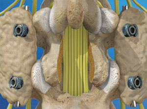
Each pedicle screw is slid over its guide wire, eased down to the vertebra, and carefully screwed into the bone.
Placing the Rods
Metal rods connect the screws to stabilize the spine. The Sextant system lets the surgeon place these rods without a large incision: a guide is positioned, and one at a time each rod is attached to the device, passed through a small incision, and seated in the screw heads, locking the screws together.
After Surgery
The instruments are removed and the incisions are closed. Most patients stay in the hospital for one to three days, as determined by the surgeon, who will also guide recovery. Because the incisions are small, healing may be quicker than after open surgery.
Questions about Lumbar Pedicle Screw Fixation (CD Horizon® Sextant®)? Call Dr. Auerbach’s office at (718) 518-5814 or send a message.
This information is for general education and does not replace advice from your doctor.
Printed from https://drauerbach.com/patient-education/lumbar-pedicle-screw-fixation-cd-horizon-sextant.html
 2:04
2:04 1:40
1:40 1:41
1:41 1:34
1:34 2:10
2:10 2:08
2:08